Super-resolution microscopy — beating the diffraction limit
An extension of the topic of diffraction and resolution. A classical optical microscope has its resolution limited by the diffraction of light (the Abbe limit, about 200 nm) — nearby objects merge into a single blob. Fluorescence techniques (STED, single-molecule localization) get around this limit by a different mechanism, making it possible to see individual molecules (Nobel Chemistry 2014).
I will give you the distance between two points — choose the method that resolves them (limits: Abbe ≈ 200 nm, localization ≈ 20 nm).
What just happened?
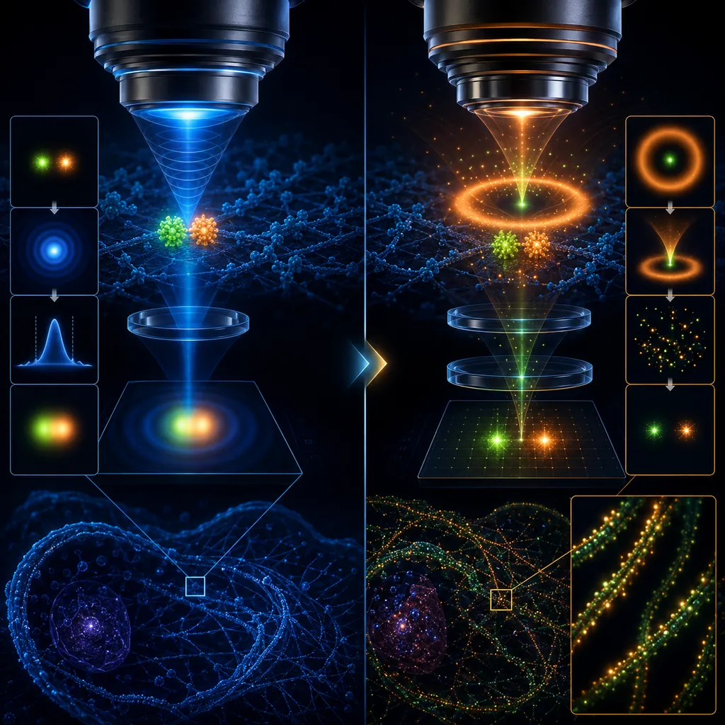
Two glowing points in the sample sit closer together than the resolution limit of a classical microscope — their images (diffraction spots) overlap and merge into a single blurred blob. Switching to the fluorescence method does not "magnify" the image — it changes the very mechanism by which the image forms, so the same two points become clearly resolved.
The Abbe limit (diffraction):
Light of wavelength λ diffracts as it passes through the objective lens — a point source never produces a perfectly sharp point in the image, but a blurred spot (the point spread function, PSF) with a width on the order of λ/2, i.e. about 200 nm for visible light. Two points closer together than d_min are indistinguishable — no matter how good the lenses are.
How does super-resolution get around this limit?
Instead of lighting up both points at once, a fluorescence technique (e.g. STORM/PALM, STED) makes only a single molecule glow at any given moment — its blurred spot can then be precisely localized (its center pinned down to within about 10–20 nm), even though the spot itself is still diffraction-sized. Repeating this thousands of times for different, randomly "blinking" molecules builds up a map of their positions — an image with a resolution far better than λ/2.
Watch out for the trap:
This is not a violation of the physics of diffraction — every single spot still has a diffraction-limited width. Super-resolution wins because it localizes the center of the spot, rather than trying to narrow it. Hence it has its own, much smaller limit — the localization precision, not the Abbe limit.
Why you need this for the exam:
Remember the relation d_min ≈ λ/2 and the association: classical microscope = diffraction limit ≈ 200 nm; super-resolution (Nobel Chemistry 2014) = getting around that limit by localizing single fluorophores, not breaking the laws of optics.
Remember: the resolution of a classical microscope is limited by the diffraction of light (d_min ≈ λ/2 ≈ 200 nm); super-resolution fluorescence microscopy gets around this limit by a different mechanism — localizing single molecules.
Theory & formulas
The diffraction limit
The resolution of a classical optical microscope is limited by the diffraction of light (the Abbe limit). Two points closer together than: merge into a single blob — for visible light this is about .
Getting around the limit
Fluorescence techniques (STED, single-molecule localization) do not break the laws of physics; they get around the limit by a different mechanism: they selectively switch the fluorescence of individual molecules on and off and precisely determine their positions.
Why it matters
It makes it possible to see and track individual proteins and processes inside living cells, for example in neurodegenerative diseases (Nobel Chemistry 2014).
Notation: — smallest distinguishable distance, — wavelength of light, — refractive index, — aperture angle of the objective.
Frequently asked questions
›What is the diffraction limit?
A physical limit on the resolution of an optical microscope, arising from the diffraction of light (≈λ/2).
›How can you "see" individual molecules?
By selectively activating/quenching the fluorescence of individual molecules and localizing them with high precision.
›Why does it matter in biology?
It lets you track individual proteins and processes inside living cells, for example in neurodegenerative diseases.
Teaching? Embed this experiment on your site.
Free widget — paste it on your school site, blog or notes. It links to the full version.